Today I am sharing a case on composite resin fillings to show the importance of radiographs in a thorough dental checkup to detect hidden decayed cavities.
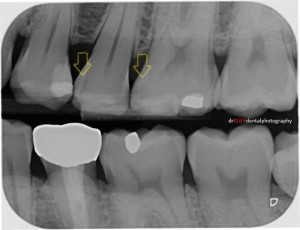
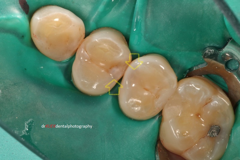
Dark shadows were detected, though challenging to the untrained eye. Decayed teeth shown (yellow arrows) on radiograph.
Mr Smith had some decay / cavities in his teeth. Although they were not obvious with a visual examination, they could be detected on the radiograph taken during his dental checkup.
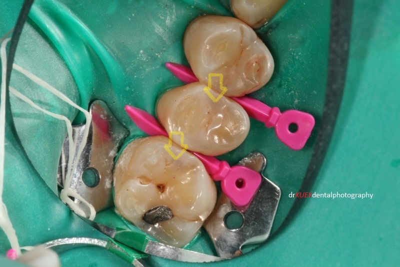
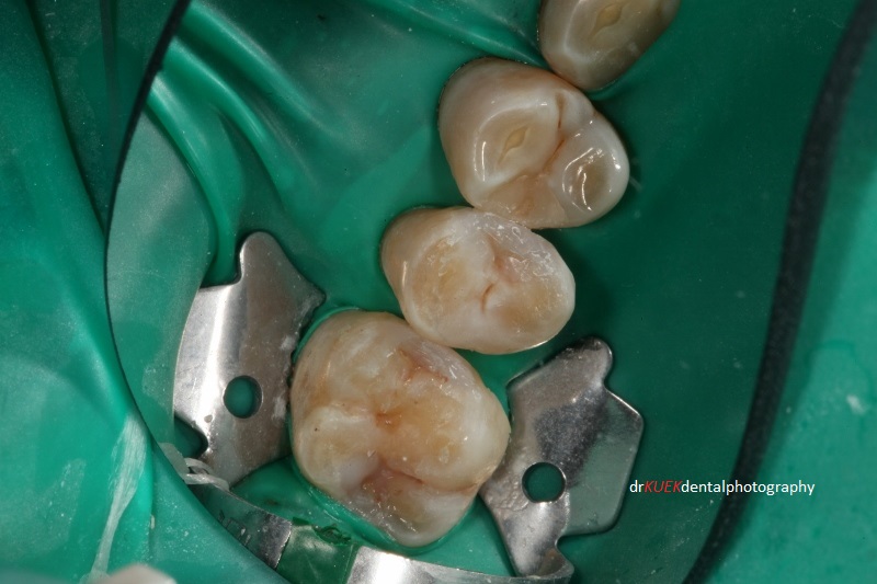
Cavities were located below the areas marked by the yellow arrows. Teeth were isolated with a rubber dam. Triodent wave-wedges (pink) were used to pre-wedge these teeth, important for good teeth contacts in the final results.
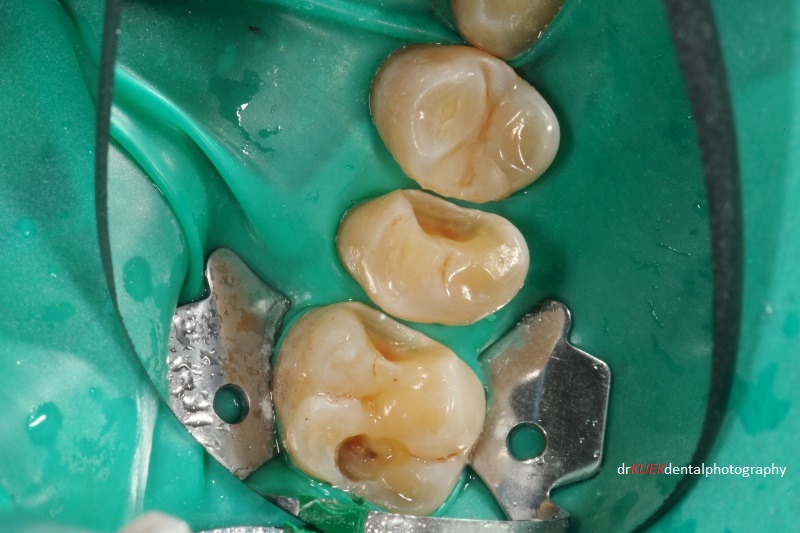
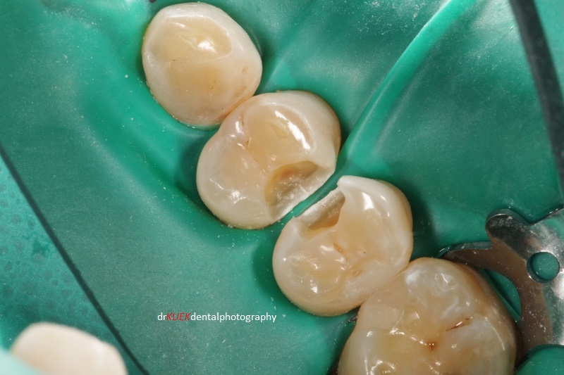
The existing amalgam filling with poor margins was removed, which revealed more decay.
The routine preparations were done to set up Mr Smith for his fillings. Once the teeth were nicely numbed, isolated and comfortable, our little project began. While I work, I always hold a mental picture of the final result that I want to achieve in terms of shape and aesthetics.
Decayed teeth areas were cleaned and inspected carefully.
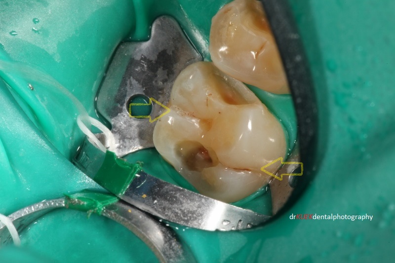
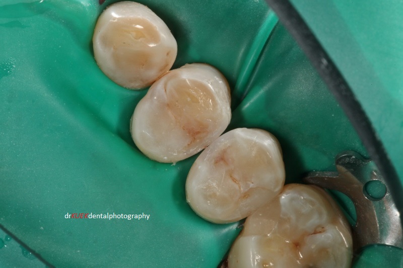
Natural grooves in this molar tooth were cleaned even though they were not decayed (yellow arrows). I then sealed these grooves to reduce the risk of future decay.
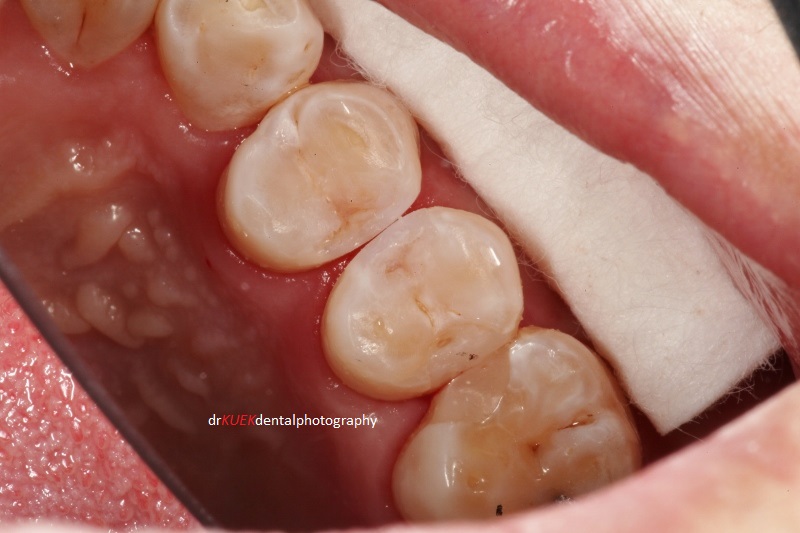
With the Triodent V3-ring and sectional matrix system (not shown – will try to in future posts), composite resin materials were carefully placed and sculpted. Placements were done in small increments and in a specific manner (I will share how it is done in the future). This is important to minimize the risk of Mr Smith feeling sensitive from these teeth later on and also ensures longer-lasting fillings. Tints were used to highlight and re-create the natural shape and aesthetics of the teeth, blending in the fillings with the natural teeth.
Shades of composite resin materials were chosen and tints used to create the fillings, aiming to blend the fillings with the natural teeth.
Mr Smith is very glad that his teeth are fixed now.
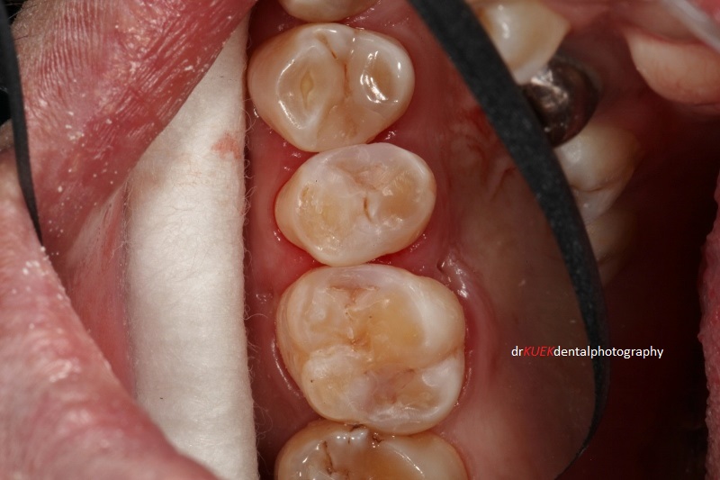
Fillings completed